
Fig. 3.
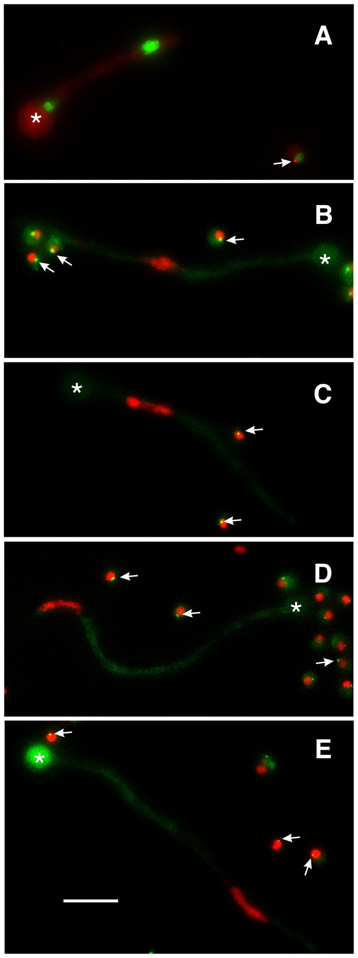
Abolition of γ-tubulin and GCP localization by deletion of gcpC. In each panel a germling carrying a gcpC deletion (gcpCΔ) is shown along with one or more ungerminated conidia that are wild-type for gcpC but carry riboB2 that prevents growth in unsupplemented media. The swollen conidia that gave rise to each germling are marked with asterisks. (A) gcpCΔ in a strain carrying histone H1-GFP and γ-tubulin-mCherry. Two nuclei of unequal sizes are present. γ-tubulin does not show SPB localization in the gcpCΔ germling, but does localize to the SPB of the nucleus in the ungerminated gcpC+ spore (arrow). (B-E) gcpCΔ in strains carrying histone H1-mCherry and GCPB-GFP (B), GCPD-GFP (C), GCPE-GFP (D) and GCPF-GFP (E). Each gcpCΔ germling has one nucleus, reflecting the fact that nuclear division is inhibited in the gcpCΔ strain. Germlings as long as these would normally have many nuclei. In each case, the GCP fails to localize to the SPB in the gcpCΔ germlings (i.e. there is no GCP spot associated with the nucleus), but localizes normally in the ungerminated gcpC+ conidia (arrows). All images are maximum intensity projections of Z-series stacks that include the entire germling so the absence of GCP fluorescence is not due to the SPB being out of the focal plane. Scale bar: 10 μm.