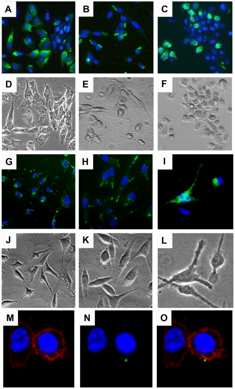
Figure 1. Binding and internalization of CPMV nanoparticles in vitro.
(A–F) Binding of CPMV to (A) BalbCl7 cells, (B) MC57 Cells, and (C) bone marrow-derived immature DCs (imDC). Cells were fixed with 2% formaldehyde and incubated with CPMV-AF488 (green) on ice overnight. The cells were washed and stained with Hoechst 33258 to visualize the nucleus (blue). (G and H) Uptake of CPMV-F by the cell lines BalbCl7 and MC57, respectively. (I) Uptake of CPMV-AF488 by imDCs. Cells were incubated overnight with the fluorescent virus particles (in green), washed with PBS, fixed, and stained with DAPI (G and H) or Hoechst 33258 (I) to visualize the nucleus (blue). (D–F and J–L) Transmission light image showing the body of the cells. The cells were visualized by fluorescence microscopy using the 20X objective. (M–O) Surface vimentin expression in Caco-2 cells stained with DAPI (blue, M–O), wheat germ agglutinin- AF555 (red, M, O), and specific antibodies to vimentin conjugated to AF647 (green, N–O). Cells were fixed and incubated with WGA one hour RT, anti-vimentin V9 antibody, 1.5 hour RT, and secondary AF647 one hour RT (M–O). Cells were visualized by fluorescence microscopy using the 60X objective (M–O).

