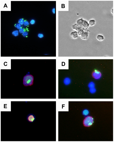
Figure 4. In vivo uptake of CPMV particles.
(A) Spleen cells were purified 4 hours after i.p. inoculation with 100 µg CPMV-AF488 and stained with DAPI. (B) Transmission light microscopy image showing the body of the spleen cells. (C–F) Localization of CPMV in splenocytes stained with: (C) PE anti-CD8α (red), (D) PE anti-CD11c (red), (E) PE anti-B220 (red), and (F) PE anti NK 1.1 (red). The cell samples were visualized under a fluorescence microscope using a 20X objective. The virus particles are in green and the nuclei are in blue.

