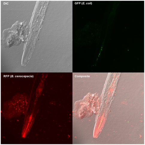
Figure 2. Localization of bacteria ingested by C. elegans.
Confocal microscopy of a live nematode that was co-cultured with E. coli DH5α, marked with green fluorescent protein, and B. cenocepacia HI2424, marked with red fluorescent protein, is shown. B. cenocepacia (red, lower left quadrant) grows throughout the nematode gut and forms aggregates on the nematode cuticle, whereas E. coli (green, upper right quadrant) is found only in limited concentration in the mouth.

