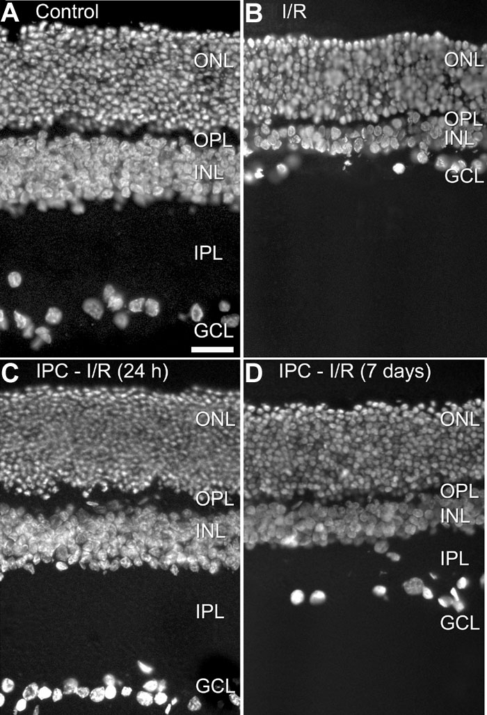
Figure 1.

Sections of rat retina stained with the fluorescent DNA-stain 4,6 diamidino 2 phenylindole (DAPI). A: Control, retina of a sham-treated eye. B: I/R, retina 7 days of reperfusion after 60 min of ischemia. Note the decrease in thickness of INL and IPL, and the reduced number of somata in the ganglion cell layer. C: IPC-I/R (24 h), retina preconditioned by 5 min of ischemia, followed 24 h later by 60 min of ischemia and sacrificed 7 days later. No histological changes can be found. D: IPC-I/R (7 days), retina preconditioned by 5 min of ischemia, followed 7 days later by 60 min of ischemia and sacrificed 7 days later. The effect of preconditioning has diminished, but the loss of IPL thickness is still partially prevented. ONL represents outer nuclear layer, OPL represents outer plexiform layer, INL represents inner nuclear layer, IPL represents inner plexiform layer, GCL represents ganglion cell layer. All images were taken at the same magnification. Scale bar in A represents 25 μm.
