Figure 4.

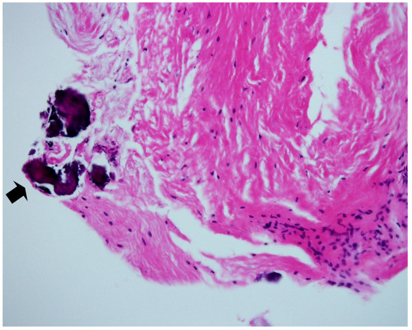
Photomicrograph of the cyst wall. The lining tissue consists of dense fibrous tissue with spotty areas of calcification (black arrow) (Hematoxylin and Eosin, ×200).

Photomicrograph of the cyst wall. The lining tissue consists of dense fibrous tissue with spotty areas of calcification (black arrow) (Hematoxylin and Eosin, ×200).