
Figure 4.
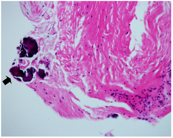
Photomicrograph of the cyst wall. The lining tissue consists of dense fibrous tissue with spotty areas of calcification (black arrow) (Hematoxylin and Eosin, ×200).
Official websites use .gov
A
.gov website belongs to an official
government organization in the United States.
Secure .gov websites use HTTPS
A lock (
) or https:// means you've safely
connected to the .gov website. Share sensitive
information only on official, secure websites.
Photomicrograph of the cyst wall. The lining tissue consists of dense fibrous tissue with spotty areas of calcification (black arrow) (Hematoxylin and Eosin, ×200).