
Figure 2.
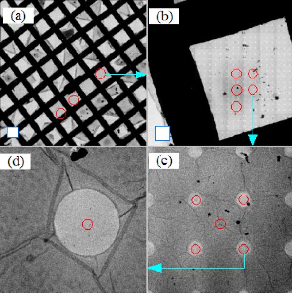
Hierarchical view of the specimen to show the process of finding the structures of interest. All images were acquired on FEI Tecnai Polara TF30 electron microscope equipped with a GATAN 4k UltraCam CCD camera. The blue square frames in the lower left corner of (a) and (b) represent the corresponding CCD imaging area, respectively. (a) Atlas map constructed at 480x nominal magnification. (b) Atlas map constructed at 3000x nominal magnification (M mode). (c) A tile image that forms the atlas map shown in (b). (d) An image taken at 14500x nominal magnification. The light blue arrows show where the location of the pointed map or images is originated in the precedent map or image.