
Fig. 8.
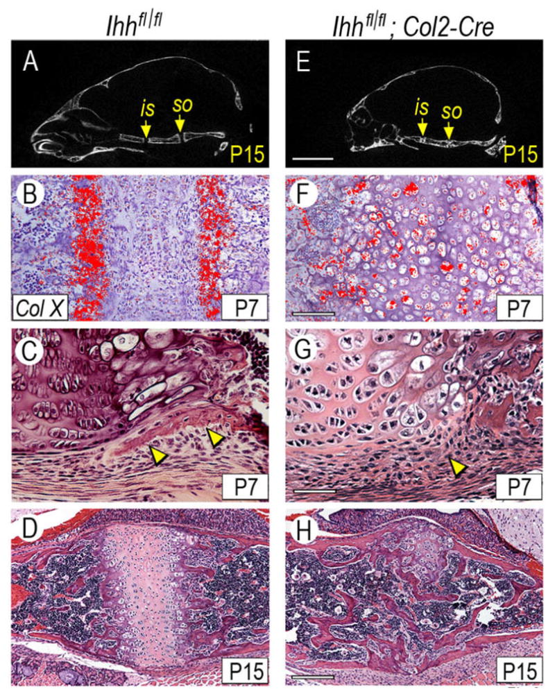
Conditional postnatal Ihh deficiency causes cranial base abnormalities. (A,E) Skulls from P15 control and Ihh-deficient mice were subjected to μCT analysis and one orthogonal plane through the cranial base is shown here. Note the presence of well defined is and so synchondroses in controls (A, arrows) and the ill-defined synchondroses and reduced antero-posterior length in mutants (E). (B–D, F–H) Sections from P7 and P15 control and mutant so synchondroses processed for collagen X gene expression (B,F) or histological analysis (C–D,G–H). Note that collagen X transcripts are restricted to hypertrophic zones in control (B) but are widespread throughout the mutant synchondrosis (F). Note also the presence of a well-formed intramembranous bone collar flanking the pre-hypertrophic and hypertrophic zones in control (C, arrowheads) that is undetectable in mutant (G, arrowhead). Note also that much of mutant synchondrosis is replaced by endochondral bone by P15 (H). Scale bar is 2 mm for A,E; 150 μm for B,F; 75 μm for C,G; and 250 μm for D,H.