
Figure 5. Caveolin-1 phosphorylation mediates H2O2-induced dissociation of VE-cadherin and β-catenin.
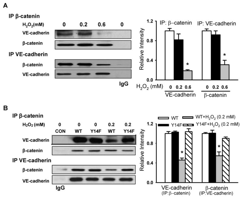
The association of VE-cadherin with β-catenin was determined by immunoprecipitation and immunoblot analysis using anti-β-catenin or anti-VE-cadherin antibodies. Left, representative Western blots for VE-cadherin and β-catenin; Right, protein quantification by densitometry. The density of proteins in each untreated (control) group was used as a standard (1 arbitrary unit) to compare the relative density in the other groups. (A) Effect of H2O2 on the association of VE-cadherin with β-catenin in naïve endothelial cells. Note dissociation upon exposure to 0.6 mmol/L H2O2. (B) Effect of H2O2 on the association of VE-cadherin with β-catenin in cells stably expressing WT and phosphorylation-defective Y14F-Cav-1 mutant. *P < 0.05, compared to control (untreated) group (A) or WT alone group (B); n = 3/each group.