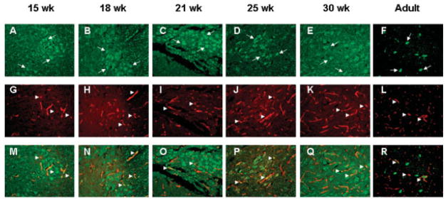
Fig 6.
Angiogenin (ANG) expression in motor neurons and blood vessels of fetal and adult human spinal cords. (A–F) Green immunofluorescence for ANG with 26-2F and Alexa 488 –labeled goat anti–mouse IgG. Arrows indicate representative ANG staining in motor neurons. (G–L) Red immunofluorescence for blood vessels with anti–von Willebrand factor polyclonal antibody and Alexa 555–labeled goat anti–rabbit IgG. Arrowheads indicate representative blood vessels. (M–R) Merge of green and red fluorescence. Arrowheads indicate colocalization of ANG and von Willebrand factor. Images are from a representative area of the ventral horns of spinal cords of (A, G, M) 15-, (B, H, N) 18-, (C, I, O) 21-, (D, J, P) 25-, and (E, K, Q) 30-week-old fetuses and an (F, L, R) adult. Original magnification ×100.

