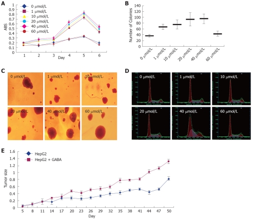
Figure 3.
Effect of GABA on the growth of HCC cells. HepG2 cells were treated with GABA at serial concentrations (0, 1, 10, 20, 40 and 60 μmol/L). A: MTT assay. Y-axis: Average value of absorbance (ABS) at 570 nm, measured with a microplate reader (n = 6, P < 0.05); B and C: Soft agar colony formation assay. Same cells were incubated in low-melting agarose as described in Materials and Methods. Two weeks later, colonies were photographed and numbers of colonies were counted. (n = 3, P < 0.05); D: Cell cycle measured by flow cytometry; E: Tumor volume in nude mice with HepG2 and GABA (0 μmol/L and 20 μmol/L) injected (mm3).

