
Figure 1.
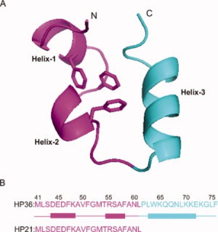
Primary structure of the villin headpiece subdomain HP36. (A) Ribbon diagram of HP36 created using PyMOL (PDB entry; 1VII). The fragment corresponding to HP21 is in purple. The three phenylalanines are shown, and the N- and C-termini are labeled. (B) Primary sequence of HP36 and HP21 with α-helices represented by rectangles. The numbering system for the full length villin headpiece subdomain is used, where the helical subdomain starts from residue Leu42. An additional Met at the N-terminus results from the expression of the protein, and this residue is designated as Met41. In the NMR structure (PDB entry 1VII), the three helices run from Asp44 to Lys48, Arg55 to Phe58, and Leu63 to Glu72, and are donated as helix-1, helix-2, and helix-3 respectively. In the X-ray structure,33 the boundaries of the helices are Asp44 to Phe51, Arg55 to Asn60, and Leu63 to Lys73.