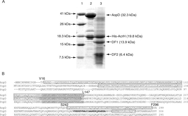
Figure 2.

Limited protease digestion of the AcrH-AopD complex. A: SDS-PAGE showing partial digestion of AcrH-AopD with chymotrypsin. Lane (1) Mw marker; lane (2) monomeric AcrH-AopD from gel filtration before digestion; and lane (3) AopD is being digested into two separate fragments, DF1 (residues 16–147) and DF2 (residues 242–296), whereas AcrH remains intact. B: Sequence alignment of AopD from Aeromonas hydrophila (AAR26342) with YopD from Yersinia enterolitica (AAD16812) and PopD from Pseudomonas aeruginosa (AAC45938). Predicted transmembrane domains (TMHMM Server v. 2.0, http://www.cbs.dtu.dk/services/TMHMM-2.0/) are highlighted in gray. Residues that are determined to be involved in chaperone binding are boxed (this work and Francis et al.32). Residues from the coiled coil regions predicted by the program COILS33 are boldfaced.
