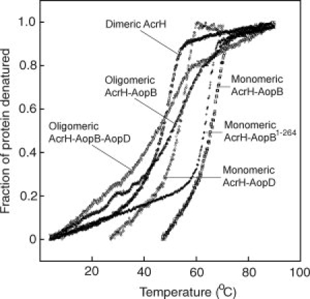
Figure 5.

Thermal denaturation of AcrH and various complexes as monitored by FarUV-CD at 222 nm. The fraction of protein denatured is plotted against temperature (°C). Legends for different proteins are: dimeric AcrH (open circle); monomeric AcrH-AopB (open square); monomeric AcrH-AopD (open rhombus); oligomeric AcrH-AopB-AopD (open inverted triangle); monomeric AcrH-AopB1–264 (cross); and oligomeric AcrH-AopB (open triangle).
