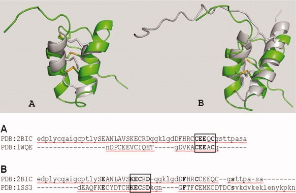
Figure 2.

PcF structural alignments. (Top) Cartoon representation of the fold alignment of PcF (PDB 2BIC) with: panel A, a representative K+-channel blocker scorpion toxin (OmTx3, PDB 1WQE) and panel B, the pollen allergen Ole e 6 from Olea europea (PDB 1SS3). Molecular rendering was performed with PyMol program. All structures are in right-left orientation starting from N-terminus. PcF is highlighted in green, whereas the other proteins are in gray. Disulphide bonds are in yellow. (Bottom) Structure-based sequence alignment was performed using SSM.13 α-helical regions are in capital letters and identical residues are in bold. Boxed: the structurally superimposed conserved residues.
