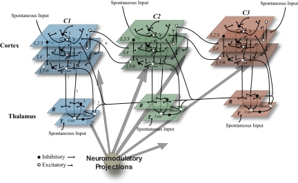
Fig. 1.
Schematic of the thalamocortical model showing 3 hierarchically organized cortical areas (C1–C3) each with an associated thalamic sector (T) and corresponding section of the reticular nucleus (R). Excitatory (white) and inhibitory (black) cells receive spontaneous input in the waking mode from areas representing cortical and subcortical structures. (1) Thalamic core cell thalamocortical loops: excitatory core cells in T project to the associated cortical area L4 (corresponding to cortical layer 4), to L5/6 (corresponding to cortical layers 5 and 6), and, via collaterals, to R. (2) Matrix cell thalamocortical loops: excitatory matrix cells in T project to L2/3 (corresponding to cortical layers 2 and 3) and L5/6 of all cortical areas as well as R. (3) Reticular nucleus network: R neurons send inhibitory projections to the neurons in the associated T sector. (4) Cortical interlaminar (vertical) connections: columnar connections are made from L4 to L2/3, L2/3 to L5/6, and L5/6 to L4 and L2/3. (5) Cortical intralaminar (horizontal) connections: each layer contains excitatory and inhibitory projections to other cells within the same layer. (6) Interareal corticocortical loops: forward projections (C1 to C2 and C2 to C3) are from excitatory L2/3 cells to L4, whereas back projections (C3 to C2 and C2 to C1) are from excitatory L5/6 cells to L2/3 and L5/6. (7) Corticothalamic connections: excitatory cells in L5/6 project via slow connections to core cells in T, forming collaterals with R, and via fast projections to matrix cells in T. (8) Diffuse neuromodulatory (cholinergic, noradrenergic, histaminergic, etc) systems project throughout the entire thalamocortical network. Not drawn to scale.

