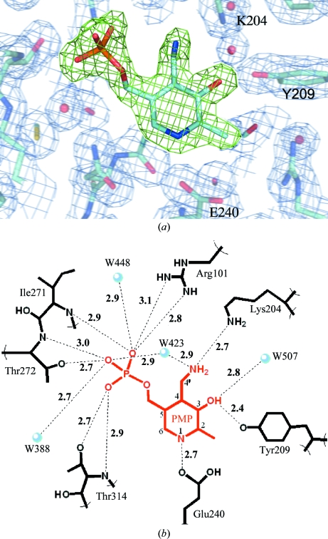
Figure 2.
(a) The PMP molecule bound in the active site is displayed using green F o − F c OMIT density contoured at 4σ (0.26 e Å−3). The surrounding active site is shown using blue 2F 0 − F c electron density contoured at 1σ (0.28 e Å−3) and a few important active-site residues are labeled. (b) The hydrogen-bonding network of PMP within the active site. PMP atoms are numbered according to convention.

