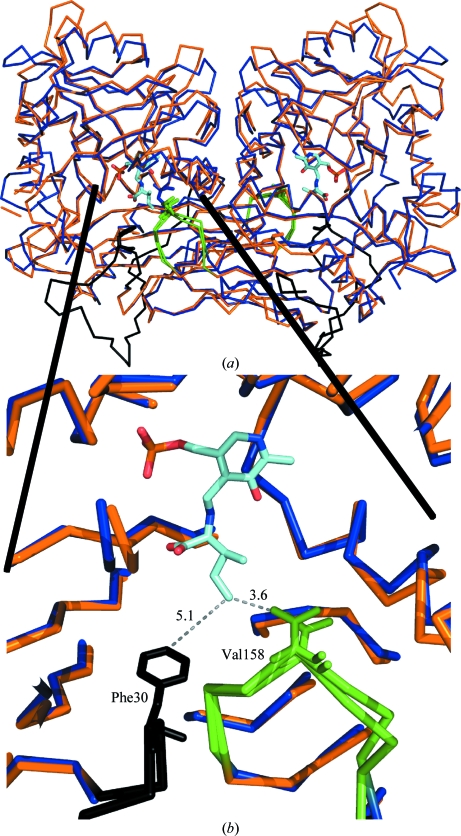
Figure 3.
Structural alignment of the MtIlvE (blue) and hBCATm (PDB code 1kt8; orange) dimers. The green loop is donated from the partner molecule and contributes a conserved valine which stabilizes the external ketimine in the active site of PDB entry 1kt8. The N-terminus of the 1kt8 structure (black loop) shows the conserved phenylalanine residue shielding the active site from bulk water molecules.

