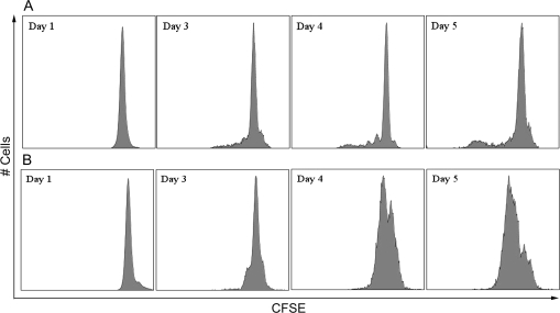
FIG. 4.
CD40L-induced proliferation of mouse and human B cells. (A) Mouse B cells or (B) human CD19+CD27− naive B cells were labeled with CFSE and then cocultured at 1.5 × 10⁶ cell/culture (mouse) or 1.5 × 10⁵ cell/culture (human) with irradiated CD40L-L cells (mouse: 5 × 10⁴ cell/culture; human: 1.5 × 10³ cell/culture) in the presence of recombinant mouse or human IL-2, IL-6, and IL-10 for up to 5 days, and CD40L stimulation was removed on day 3 (mouse) or day 4 (human). Cells were harvested at the indicated time points and assessed by flow cytometry. Dead cells were identified by staining with Live/Dead near-infrared (mouse) or red (human) Staining Kit and excluded from data analysis. These data are representative of two separate experiments (for humans, experiment used B cells from one individual donor) with three replicates per time point.

