Figure 6.
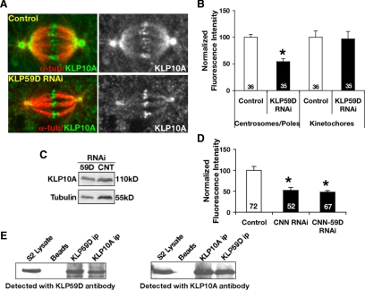
KLP59D targets KLP10A to centrosomes/spindle poles. (A) Representative examples of RNAi-treated cells fixed, immunostained with anti-KLP10A (green) and anti-α-tubulin (red), and imaged using identical methods (left). Right panel, only KLP10A labeling. (B) Average KLP10A immunofluorescence intensities at spindle poles/centrosomes and kinetochores. Values are normalized so that control RNAi intensities are 100%. KLP10A levels decrease ∼50% upon KLP59D RNAi; p < 0.001. (C) Western blots of KLP59D RNAi cell lysate demonstrate a slight increase (1.3-fold) in total KLP10A compared with control (CNT). (D) Levels of pole-associated KLP10A in cells treated with centrosomin (CNN) RNAi or co-RNAi with KLP59D. Values are normalized so that control intensities are 100%. (E) Western blots of cross-immunoprecipitations of KLP59D and KLP10A from S2 cell lysates. Beads, protein G beads only; ip, immunoprecipitate.

