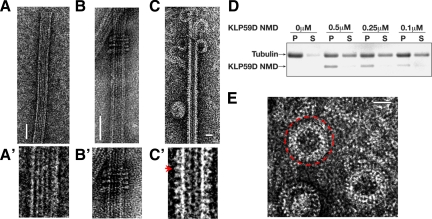
Figure 7.
Electron microscopy analysis of KLP59D. (A) Electron micrographs of negatively stained MTs. (A′) Higher magnification of A. (B) KLP10A NMD forms MT associated rings. (B′) Higher magnification of B. (C) KLP59D neck/motor domain (NMD) decorates the MT lattice and generates protofilament “peels” at MT ends in the presence of AMPPNP. (C′) Higher magnification of B. KLP59D motors visible as the small protrusions from the MT lattice (marked with red arrow). (D) KLP59D disassembles MTs in vitro. Taxol-stabilized MTs were incubated with the indicated concentrations of purified KLP59D NMD in the presence of ATP. After centrifugation, pellet (P) and supernatant (S) samples were analyzed by SDS-PAGE and Coomassie-stained. (E) Enlargement of isolated rings; one ring is marked with a red circle. Scale bars, 50 nm.

