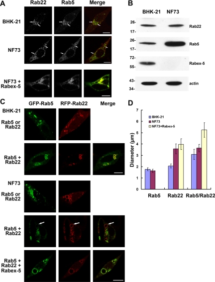
Figure 5.
Functional synergy of Rab22 and Rab5 on early endosomes requires Rabex-5. A. Confocal fluorescence microscopy showing colocalization of endogenous Rab22 and Rab5 in BHK and NF73 cells. NF73 cells were either observed directly or after expression of Rabex-5 as indicated. The cells were fixed, permeabilized, and immunostained with an anti-Rab22 rabbit polyclonal antibody and an anti-Rab5 mAb, followed by confocal fluorescence microscopy. The individual channels (green and red) are shown in black and white, whereas the merged images are shown in color. Bar, 8 μm. (B) Immunoblot showing endogenous levels of Rab5, Rab22, and Rabex-5 in BHK and NF73 cells. Cell lysates were subjected to SDS-PAGE, followed by immunoblot analysis with the anti-Rab5, anti-Rab22, and anti-Rabex-5 antibodies as indicated. The same membrane was also probed with the anti-actin antibody, as a loading control. Molecular mass standards (in kilodaltons) are indicated on the left side of the panels. The results were reproducible in two independent experiments. (C) Confocal fluorescence microscopy showing synergistic enlargement of early endosomes by GFP-Rab5 and RFP-Rab22 in BHK cells but not in Rabex-5–deficient NF73 cells. GFP-Rab5 and RFP-Rab22 were expressed either separately or together in the cells, as indicated, followed by confocal fluorescence microscopy. The coexpression of Myc-Rabex-5 in the bottom panel was confirmed by immunofluorescence microscopy (data not shown). The results were reproducible in three independent experiments. Bar, 8 μm. D. Quantification of early endosomal size in BHK and NF73 cells. The graph shows the maximal size of early endosomes labeled by GFP-Rab5 alone, RFP-Rab22 alone, or both of the Rabs. In each case, the diameters of 100 of the largest endosomes in ∼40 cells were measured and shown are the mean and calculated SEM.

