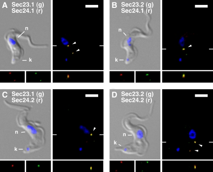
Figure 6.
All TbSec23 and TbSec24 subunits colocalize in T. brucei. As indicated (A–D) the doubly HA/Ty epitope-tagged cell lines were fixed, permeabilized, and immunostained with anti-HA (staining TbSec23.1:HA or TbSec23.2:HA) and anti-Ty (staining TbSec24.1:Ty or TbSec24.2:Ty). After staining with appropriate secondary antibodies (anti-HA, green and anti-Ty, red), cells were imaged by epifluorescence microscopy. For each pair, merged DAPI/DIC images with stained nucleus (n) and kinetoplast (k) indicated are presented (top left). Corresponding three-channel summed stack projections are presented (top right) with discrete sites (1 or 2 per cell) at which each pair of TbSec subunits colocalize indicated (arrowheads). Single channel red and green images in the region of colocalization are presented (bottom left), as are images of the x-z transect through the major region of colocalization (bottom right). The planes of the x-z transects are indicated by marginal hatch marks in the three channel x-y images. Bars, 5 μm.

