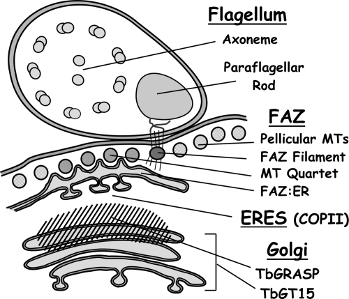
Figure 9.
Diagram of ERES:Golgi junctions in BSF trypanosomes. A partial cross section of a cell in the postnuclear region transecting the junction of an ERES and Golgi complex is represented. Major structures include the flagellum containing the 9 + 2 axoneme and paraflagellar rod; the FAZ, including pellicular microtubules, FAZ filament, antiparallel microtubule quartet, and the FAZ:ER; the ERES defined by COPII subunits; and the Golgi defined by TbGT15 and TbGRASP. Diagram inspired by Bastin et al., 2000 and Hill, 2003).

