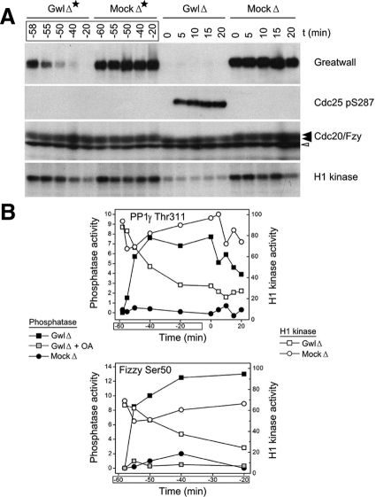
Figure 4.
When Gwl is depleted from CSF extracts, phosphatase activity is induced before MPF activity is lost. (A) Protein A beads coated with purified anti-Gwl antibodies, or control protein A beads, were added at t = −60 min to CSF extracts. To investigate the state of the extracts during the course of the immunodepletion, aliquots were taken at the indicated times (GwlΔ* and MockΔ*). At t = 0, extracts that had been immunodepleted for 1 h at 4°C were then incubated at 22°C and processed as in Figure 1 (i.e., GwlΔ* and GwlΔ indicate successive examinations of the same sample, the former during the immunodepletion at 4°C and the latter after transfer of the depleted samples to 22°C). (B) The samples in A were also analyzed for phosphatase activity against the CDK phosphosites at Thr311 of PP1γ (top) and Ser50 of Cdc20/Fizzy (bottom). The results are plotted on the same graph with quantitative data from phosphorimager scans of the H1 kinase assays shown in A. Note that the GwlΔ* samples from t = −55 min and t = −50 min reveal the induction of considerable phosphatase activity even though H1 kinase (i.e., CDK) activity remains at high, M phase-like levels similar to those in the mock depletion.

