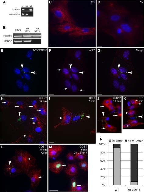
Figure 4.
Disrupting the CENP-F/Hook2 interaction interferes with MT repolymerization after nocodazole challenge. (A) WT and CENP-F−/− MEFs were isolated and genotyped. The top bands use PCR primers flanking the 5′ loxP site of the floxed CENP-F allele, determining floxed versus WT. The bottom bands use PCR primers flanking the entire floxed sequence, allowing effective PCR only once the internal sequence has been excised, thus “recombination” has occurred. (B) RT-PCR confirmation of CENP-F−/− MEFs. NDRG4 primers and C2C12 lysate were used as positive controls. (C–D) WT and CENP-F−/− cells were treated with nocodazole, washed with fresh medium, and fixed after 5 min. (C) WT MEFs display reconstituted MT networks whereas (D) CENP-F−/− MEFs show absence of MT repolymerization. MTs are labeled in red. (E–G) Expression of NT-CENP-F (green) displaces endogenous Hook2 (red) expression from centrosome (arrowheads). Arrows indicate typical Hook2 localization to centrosome in untransfected cells. (H–M) Four cell lines were transfected with NT-CENP-F-GFP, treated with nocodazole for 2 h, washed with media, and allowed to recover. Transfected constructs are labeled green with an arrow, MTs are labeled red, and nuclei are labeled blue with 4,6-diamidino-2-phenylindole. (H–L) Arrows indicate MTOC aster formation in untransfected cells, whereas arrowheads point to NT-CENP-F–expressing cells without MTOC asters. (H) COS-7 cells at 5 min after washout (I) HeLa cells at 5 min. (J) C2C12 cells at 10 min. (K) 3T3 cells at 20 min. (L) Cold-treatment phenocopies nocodazole treatment in COS-7 cells. (M) Negative control C-terminal CENP-F construct (arrow) does not have the same effect, showing the specificity of this phenotype. (N) Quantification of MTOC aster formation in COS-7 cells after 5 min. Gray indicates percentage of counted cells with a MTOC aster, whereas black represents the percentage with no MTOC aster. WT cells reconstitute MTOC asters in >90% of cells, whereas NT-CENP-F–expressing cells repolymerize <10% MTOC asters. P < 0.0001. Bars, 10 μm.

