Figure 7.
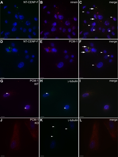
Centrosomal proteins are redistributed by CENP-F disruption and deletion. (A–F) COS-7 cells expressing NT-CENP-F show colocalization of the CENP-F truncation puncta with ninein and PCM-1. An arrowhead indicates normal centrosomal protein expression and aberrant colocalization with NT-CENP-F is labeled with an arrow. (A) NT-CENP-F. (B) ninein. (C) Merge. (D) NT-CENP-F. (E) PCM-1. (F) Merge. (G–L) WT and CENP-F−/− MEFs were labeled for PCM-1 (red) and γ-tubulin (green). (I) PCM-1 clearly localizes with γ-tubulin in WT MEFs, whereas in (L) CENP-F−/− MEFs, PCM-1 is diffusely cytoplasmic. Bars, 10 μm.

