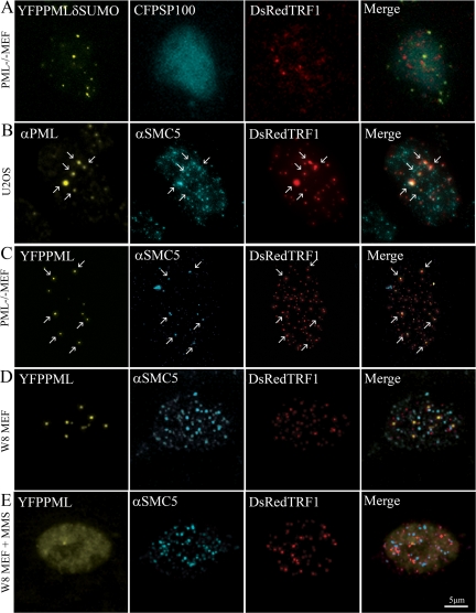
Figure 9.
A role for SUMO in the formation of new PML bodies. (A) SUMOylation-deficient PML does not localize at telomeric DNA in PML−/− MEFs. PML−/− MEFs were cotransfected with DsRed-TRF1, ECFP-Sp100 and YFP-tagged SUMOylation-deficient PML. The image shows the formation of PML aggregates (yellow), which do not colocalize with Sp100 (cyan) and do not localize at telomeric DNA that are stained by DsRed-TRF1 (red). (B) The SMC5/6 complex is present at sites where PML bodies form at telomeric DNA. U2OS cells were transfected with DsRed-TRF1, treated with MMS, and allowed to recover in fresh medium. Cells were fixed and stained with anti-PML and anti-SMC5 antibodies. The arrows point to positions in the nucleus where PML (yellow) and SMC5 (cyan) localize at telomeric sites (red). (C) Ectopically expressed PML localizes together with SMC5 at telomeric DNA in PML−/− MEFs. PML−/− MEFs were cotransfected with EYFP-PML, to induce PML body formation, and DsRed-TRF1, fixed, and stained with anti-SMC5 antibody. The arrows indicate the positions where PML (yellow) and SMC5 (cyan) localize at telomeric DNA (red). (D) PML and SMC5 do not localize at telomeric DNA in untreated and (E) MMS-treated W8 MEFs. W8 MEFs were cotransfected with YFP-PML and DsRed-TRF1 and either not treated or treated with MMS. Cells were fixed (without recovery) and stained with anti-SMC5 antibody. Both untreated and MMS-treated cells show that the SMC5/6 complex (cyan) localize in a punctate/diffuse pattern, which does not colocalize with telomeric sites (red) or PML (yellow).

