Figure 1.
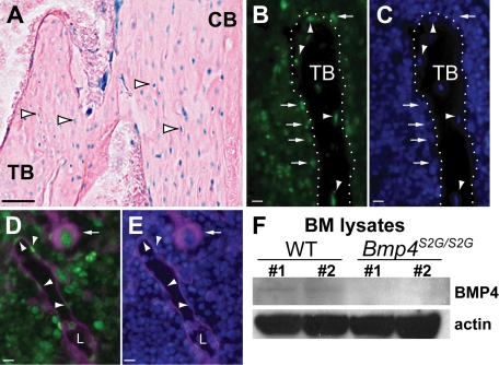
BMP4 is expressed in hematopoietic niches and BMP4 expression is reduced in the BM of Bmp4S2G/S2G mice. (A-E) Analysis of Bmp4 expression in the long bones in Bmp4lacZ/+ reporter mice. (A) β-Galactosidase enzyme activity (blue) is detected in the nuclei of trabecular (TB) and cortical (CB) bone-encased osteocytes (arrowheads), bone-lining cells, and BM cells. (B-C) Staining with (B) anti–β-gal antibody (green) and (C) DAPI (blue) confirms Bmp4 is expressed in osteocytes (arrowheads) and bone-lining osteoblasts (arrows). Trabecular bone (TB) is outlined by the dotted white line. (D-E) CD31 and β-gal colocalize in the BM. (D) Merged image showing endothelial cells (arrowheads) and megakaryocytes (arrow) are among the BM cells costained with β-gal (green) and CD31 (magenta). L indicates blood vessel lumen. (E) Merged image showing DAPI (blue) and CD31 (magenta) expression. (F) Western analysis shows reduced levels of mature BMP4 (∼ 24 kDa) expression in the BM of Bmp4S2G/S2G mice (n = 2) compared with WT controls (n = 2). Actin (∼ 42 kDa) expression was probed as a loading control. Scale bars: (A) 80 μm, (B-E) 20 μm. See “In situ BMP4 expression analysis” for details about image acquisition and processing.

