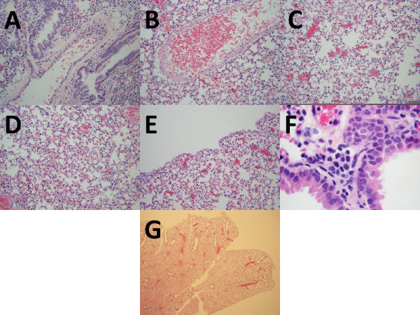
Figure 2.
Photomicrographs of control mouse lungs. a. Bronchoarterial space without inflammation (hematoxylin and eosin, original maginification 200 ×). b. Pulmonary vein without inflammation (hematoxylin and eosin, original maginification 200 ×); c. Amuscular blood vessels without inflammation (hematoxylin and eosin, original maginification 200 ×); d. Inter-alveolar spaces without inflammation (hematoxylin and eosin, original maginification 200 ×); e. Pleura lacking inflammation. (hematoxylin and eosin, original maginification 200 ×); f. Small peri-bronchial inflammatory aggregate without eosinophils (hematoxylin and eosin, original maginification 1000 ×); g. Low power appearance (hematoxylin and eosin, original magnification 40 ×).

