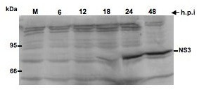
Figure 2.

Kinetics of BVDV NS3 protein expression in infected cells. MDBK cells were infected with BVDV at an MOI of 5 and the cell lysates prepared at the indicated time points. Rabbit anti-NS3 polyclonal antibody was used at 1/1000 dilution. Goat anti-rabbit alkaline phosphatase-conjugated secondary antibody was used to detect NS3 protein. M: Mock-infected cell lysate collected at 24 h after incubation. Higher NS3 expression levels are seen at 24 h and 48 h.p.i.
