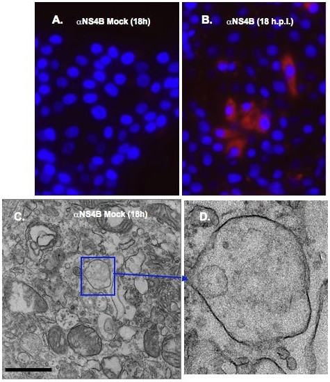
Figure 7.

Immunostaining of BVDV-infected MDBK cells. Cells were plated in 8-chamber slides, mock infected or infected with BVDV. At 18 h.p.i, cells were fixed with 4% formaldehyde/0.1% glutaraldehyde for 10 min. Cells were permeabilized with 0.05% Triton X-100, stained with NS4B-specific antibody and Qdots 605-conjugated secondary antibody (Molecular Probes, Invitrogen, CA), followed by fluorescence microscopy. Nuclei were stained with DAPI. Notice the red stain in BVDV-infected cells (B) and no stain in mock-infected cells (A). Labeled cells were fixed with 2.5% glutaraldehyde prior to sectioning and TEM analysis. Boxed area indicates the vesicular structures in mock-infected cells (C). A higher magnification of the boxed area is shown in (D). No electron dense Qdots were observed in mock-infected cells (D). Bars = 1 μm.
