
Figure 8.
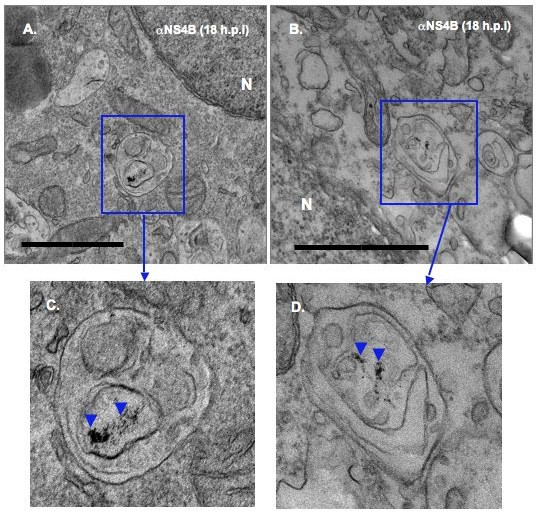
Immunodetection of NS4B protein in BVDV-induced membranes. BVDV-infected cells were processed as above for ultrastructural analysis. Notice the presence of electron dense Qdots in vesicular structures from BVDV-infected cells [rectangle areas in (A) and (B); arrowheads in (C) and (D)]. Higher magnifications of the boxed areas are shown in (C) and (D). Bars = 1 μm.