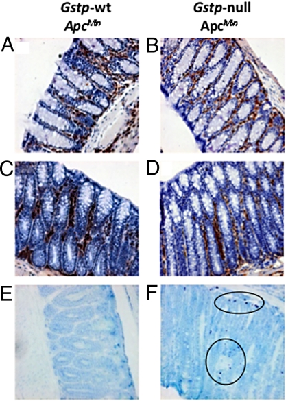
Fig. 5.
Increased mast cell infiltrates in the Gstp-null ApcMin colon. Immunohistochemical staining of (A, C, and E) Gstp-wt ApcMin and (B, D, and F) Gstp-null ApcMin was carried out to detect immune cell infiltrates. Tissues from both genotypes were found to have substantial infiltration of macrophages (A and B, magnification ×200) and large numbers of neutrophils were also present in both genotypes (C and D, magnification ×200). Greatly increased numbers of mast cell infiltrates were found in the colon of (F) Gstp-null ApcMin mice relative to (E) Gstp-wt ApcMin mice (circles indicate mast cells; magnification ×200). For further details, see Materials and Methods.

