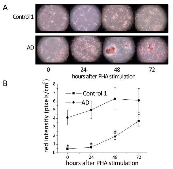
Figure 2.
Neutral lipid accumulation in PHA-stimulated PBMCs from control 1 subjects and AD patients. Immediately after separation, cells were incubated with PHA for the time periods indicated. After harvesting, cells were washed, fixed by soaking in 10% formalin, stained with ORO for NL, and counter-stained with Mayer's hematoxylin for nuclei. Cells were then examined by light microscopy and two different fields per sample were imaged. Red ORO intensity was measured in these two fields using NIH Image J software. Panel A shows ORO staining images for one control 1 (84 years old; ORO score 0 at 0 time) and one AD (85 years old; ORO score 3 at 0 time), which are representative of about thirty subjects for each group. Panel B shows red intensity expressed as mean pixels ± SE/cm2, as described in Materials and Methods. *P < 0.01, compared with control 1 by Student's t test.

