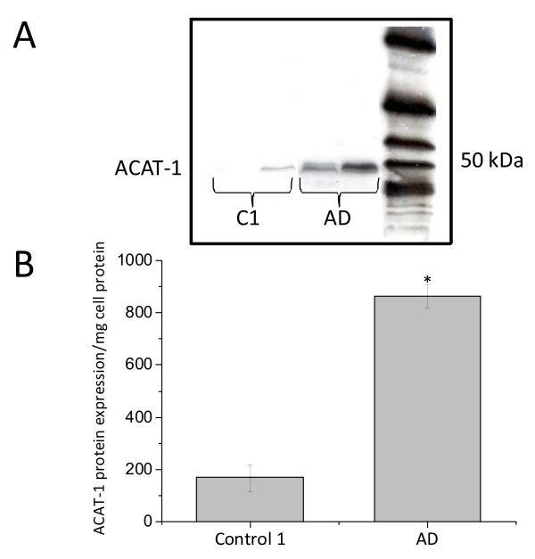
Figure 5.
ACAT-1 protein expression in control 1 and AD PBMCs. Western blotting with DM10 was used to monitor ACAT-1 protein content. The anti-ACAT-1 DM10 antibody specifically recognized a single protein band from PBMC extracts with an apparent molecular weight of about 50 kDa. No other protein signal(s) was detectable. A. Representative ACAT-1 immunoblot. B. Results of densitometric analysis by Scion Image software of ACAT-1 protein immunoreactivity relative to β-actin in 20 AD patients and 20 control 1. Data values are represented as mean ± SE *P = 0.000 vs control 1.

