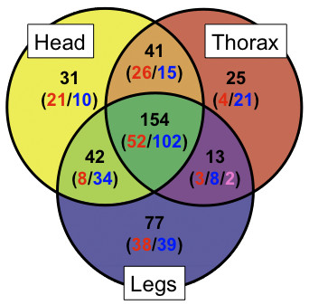
Figure 3.

Expression differences between horn and leg primordia relative to abdominal epithelium. Categorization of genes that exhibited significantly differential (p value < 0.05 and > 2-fold difference). The labels on each category represent the tissue types (head = head horns, thorax = prothoracic horns, and legs = legs). Numbers indicated in the Venn diagram represent the counts of non-redundant sequences in each category. The numbers in parenthesis indicate the counts of sequences that showed enriched or depleted expression relative to abdominal epithelium, where: red = enriched, blue = depleted, and pink = mixed (i.e. enriched in thoracic horns and depleted in legs).
