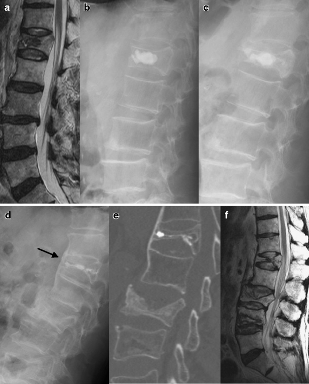
Fig. 3.

Radiologic studies of an 80-year-old man with an L1 compression fracture. a The initial MRI showed an acute compression fracture with osteonecrosis in the L1 vertebral body. b Immediate postoperative lateral plain X-ray showed well-deposited CaP cement. c Six months after the vertebroplasty, recollapse and heterotopic ossification occurred. The lateral plain X-ray (d), computed tomography (e) and MRI (f) were taken after 26 months after the vertebroplasty. The injected CaP was reabsorbed. Heterotopic ossification progressed and bone fusion developed (arrow). A subsequent vertebral compression fracture occurred at the L3 and L4 vertebrae
