
Fig. 1.
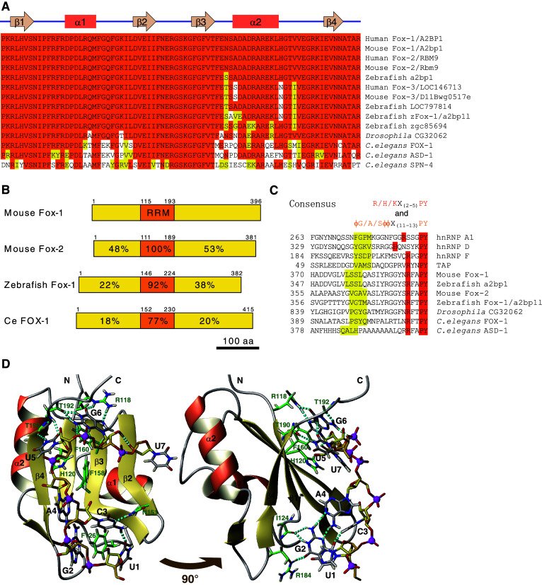
The Fox-1 family RNA-binding proteins are phylogenetically conserved. a Amino acid sequence alignment of the RRM domain of the Fox-1 family proteins in human and mouse (Fox-1, Fox-2, and Fox-3), zebrafish (a2bp1, LOC797814, zFox-1/2bp1l, and zgc85694), fruitfly (Drosophila CG32062), and nematode (C. elegans FOX-1, ASD-1, and SPN-4). Identical amino acid residues are shaded in orange and residues with similar properties are in yellow. The secondary structure of the human Fox-1 RRM domain is indicated above the alignment [23]. b Schematic illustration of the domain structure of mouse Fox-1 and Fox-2, zebrafish Fox-1/A2bp1l, and C. elegans FOX-1. RRM domain is in orange. Identities of amino acid sequences of each domain compared to that of mouse Fox-1 are indicated. Amino acid positions are indicated. c Amino acid sequence alignment of putative hPY-NLS of the Fox-1 family proteins. The C-terminal-most regions of mouse Fox-1, Fox-2, zebrafish a2bp1, zFox-1/2bp1l, Drosophila CG32062, and C. elegans FOX-1 and ASD-1 are aligned with confirmed hPY-NLS of human hnRNP A1, hnRNP D, hnRNP F, and TAP [18]. Residues that match the consensus of hPY-NLS [18] are shaded in orange and yellow. ϕ, hydrophobic side chain. Positions of the N-terminal-most residues are indicated. d Solution structure of the RRM domain of Fox-1/Fox-2 in complex with 5′-UGCAUGU-3′ in ribbon (protein backbone) and stick (RNA) representation. Right panel Image of left panel rotated by 90° as indicated. Important protein side chains and peptide bonds involved in hydrophobic or static interactions with the RNA are represented as sticks. Oxygen, nitrogen, hydrogen, and phosphorus atoms are in red, blue, light yellow, and magenta, respectively. Carbon atoms in the protein, the RNA backbone, and bases are in green, yellow, and gray, respectively. Hydrogen bonds are indicated as cyan dotted sticks. Hydrogen atoms in the RNA backbone are omitted except for one involved in a hydrogen bond. Illustrations were generated from a dataset deposited as ‘2err’ in PDBj database with MOLMOL [78]