Figure 2.
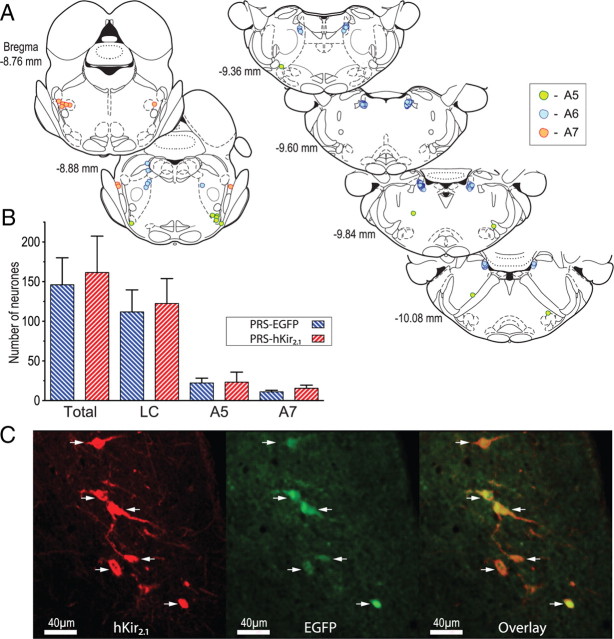
Retrograde transduction of pontospinal NAergic neurons by adenoviral vectors. A, Schematic showing the typical pattern of retrograde labeling of NAergic somata in the pons after bilateral AVV injection in the lumbar dorsal horn (taken from a single representative animal). The majority of neurons were located in the ventral LC (blue symbols) with a cluster in A7 (red) and a smattering of neurons in the A5 region (green). (Neurons plotted on sections from Paxinos and Watson, 2005). B, AVV-PRS-hKir2.1 transduced similar numbers of neurons as AVV-PRS-EGFP (n = 4 rats/group), with a majority found in the LC (76%), and the remainder found in A5 (14%) or A7 (10%). C, After dorsal horn injection of AVV-PRS-hKir2.1 in vivo, IHC for hKir2.1 showed labeling in a subset of NAergic neurons (shown here in ventral LC). The same subset of NA neurons also showed EGFP fluorescence confirming that they had been retrogradely transduced by the AVV. The overlaid EGFP and hKir2.1 images illustrates that hKir2.1 expression was restricted to EGFP-positive neurons (arrows) and there was no evidence of hKir2.1 expression in the surrounding LC neurons.

