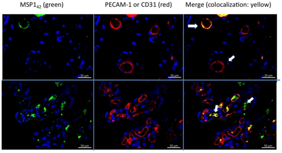
Figure 7. MSP142 co-localizes with fetal endothelial cells following perfusion.
MSP142 (green staining) co-localizes with CD31 (PECAM-1, red staining) indicating its presence in and around the fetal vascular endothelial cells (upper and lower panels for placentas 1 and 5 respectively). Similar co-localization was observed for placentas 2 and 3 (not shown). The upper panels are at 400× and lower at 200× magnification.

