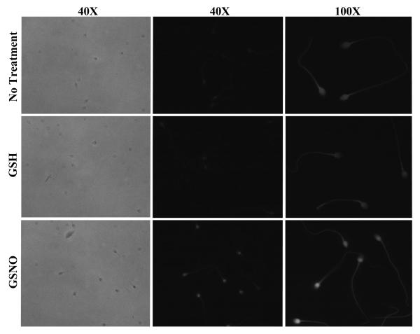
Figure 2. In situ detection of S-nitrosylated targets in human spermatozoa.
Spermatozoa were incubated without (no treatment; top) or with GSH (middle) or GSNO (bottom) for 1 hr prior S-nitrosylated proteins labelling with Texas-red fluorescence-MTSEA as described in Materials and Methods. Left and middle columns represent phase and fluorescence images, respectively, using the 40X objective. On the right column, a more detailed localisation of sperm S-nitrosylation labelling is shown using 100X objective. Results of 1 experiment presented are representative of 6 others performed with different sperm donors.

