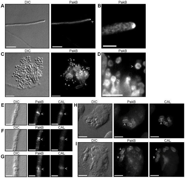
Figure 2. PakB localization during vegetative growth and asexual development at 25°C and during macrophage infection at 37°C.
pakB+ HA strains were grown on ANM + (NH4)2SO4 for 4 days at 25°C (A–G) or in LPS activated J774 murine macrophages for 24 hours (H–I) and immunofluorescently labeled with 3F10 rat monoclonal anti-HA primary and an ALEXA 488 goat anti-rat secondary antibody (PakB) and stained with calcofluor (CAL). (A) PakB is concentrated at the hyphal apex, indicated by the white arrowhead. (B) Magnification of region indicated by arrowhead in (A). (C) PakB is localized throughout all of the cell types of the conidiophore; metulae, phialides and newly formed conidia. (D) Magnification of region indicated by the white arrowhead in (C). PakB is concentrated at the phialide to conidium interface (black arrowhead) and around the periphery of newly formed conidia (white arrowhead). PakB is not visible in older conidia. (E–G) PakB is localized to nascent septation sites where it is co-localized with calcofluor stained septa, as a single band (E), as two bands on either side of the calcofluor stained septa (F) or as two spots on either side of the calcofluor stained septa (G). (H–I) During macrophage infection, PakB is localized around the yeast cell periphery. (H) PakB is not observed localized either at nascent septation sites prior to, or immediately after (white arrowheads), cell wall deposition. (I) Localization at septation sites can be observed prior to cell separation in vivo (white arrowheads). PakB is localized to the division site and to the region immediately adjacent during cell separation in vivo (double white arrowheads). Scale bars, 20 µm (A,C, H–I), 10 µm (D) and 2.5 µm (B, E–G).

