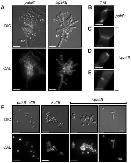
Figure 7. ΔpakB conidiophores display septation defects.
Wildtype (pakB +), ΔcflB and ΔpakB strains were grown on ANM + (NH4)2SO4 for 14 days at 25°C. (A) Conidiophores of wildtype (pakB +) and ΔpakB strains. The conidiophores are comprised of a stalk (s), metulae (m), phialides (p) and chains of conidia (c). All cell types are observed in the ΔpakB strains. Conidia, and occasionally phialides, of ΔpakB conidiophores are misshapen and larger in size. Unlike the chains of conidia observed in wildtype conidiophores, more than one conidium per phialide is rarely observed in the ΔpakB conidiophores. (B–E) Septa in conidiophores. In wildtype conidiophores (B), two separate chitin disks can be observed at phialide to conidium and conidium to conidium connections, indicating cellular division and cell separation. This is not observed in the ΔpakB conidiophores (C–E). Only one septum (C), no septa (D) or incomplete septa (E) are observed at the phialide to conidia cell boundaries. (F) Conidia were scraped off the colony surface, re-suspended in 0.005% Tween 80 solution, filtered through Miracloth and stained with calcofluor (CAL). Conidia of wildtype (pakB+ cflB+) are uniform in size. In contrast, conidia of the ΔcflB strain varied in size. Similar to the ΔcflB strain, conidia of the ΔpakB strain also varied in size. However, conidia of the ΔpakB strain were swollen to a greater extent than those from the ΔcflB strain and often exhibited uneven calcofluor staining. In addition, numerous yeast cells were observed in the ΔpakB strain. Images were captured using differential interference contrast (DIC) or with epifluorescence to observe calcofluor stained fungal cell walls (CAL). Scale bars, 20 µm (A and F) and 2.5 µm (B–E).

