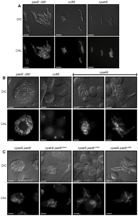
Figure 8. The ΔpakB strain displays morphological defects in vivo.
(A) Wildtype (pakB + cflB +), ΔcflB and ΔpakB strains were grown on BHI slides for 5 days at 37°C. Wildtype produces numerous yeast cells from arthroconidiating hyphae. Both the ΔcflB and ΔpakB strains produce numerous yeast cells from arthroconidiating hyphae, which are indistinguishable from wildtype. (B) LPS activated J774 murine macrophages infected with conidial suspensions of the wildtype (pakB + cflB +), ΔcflB and ΔpakB strains. After 24 hours, numerous yeast cells dividing by fission were observed in macrophages infected with wildtype (pakB + cflB +). In contrast, conidia of the ΔcflB strain remains predominately ungerminated in infected macrophages. Inside macrophages, some of the conidia of the ΔpakB strain appear large, swollen and ungerminated. However, the majority of infected macrophages contain highly branched, septate, hyphal ΔpakB cells. (C) LPS activated J774 murine macrophages infected with conidial suspensions of the ΔpakB pakB +, ΔpakB pakBH204G, ΔpakB pakB ΔCRIB and ΔpakB pakB ΔGBB strains. After 24 hrs, the yeast cells produced by the ΔpakB pakB + and ΔpakB pakBH204G strains are indistinguishable from wildtype. Similar to the ΔpakB mutant, the yeast cells produced by both the ΔpakB pakB ΔCRIB and ΔpakB pakB ΔGBB strains in vivo are long and septate. Images were captured using differential interference contrast (DIC) or with epifluorescence to observe calcofluor stained fungal cell walls (CAL). Scale bars, 20 µm.

