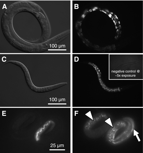
Fig. 6.
Analysis of acidic gut granules in adult worms. LysoTracker Red was used to stain acidic gut granules in 3-day-old control (A and B) and vha-6(RNAi) worms (C and D). Staining intensity varied along the length of the intestine, with posterior intestinal cells displaying the brightest signal. Control and vha-6(RNAi) worms stained similarly, despite the obvious difference in developmental age (L2 vs. young adult); developmentally matched worms similarly showed no obvious differences in the formation of acidic granules. Inset in D shows a negative control (without LysoTracker Red) that was exposed 5 times as long, demonstrating lack of background. For examination of autofluorescent gut granules, 3-fold embryos from the mosaic vha-6(ok1825) Pvha-6::VHA-6::mCherry rescue line were imaged at 555-nm excitation and 590-nm emission for transgene expression (E) and at 340-nm excitation and 535-nm emission for autofluorescence (F). Embryo at right is expressing mCherry rescue marker at the apical membrane of the lumen, which fluoresces red (E), as well as a muscle Pmyo-3::GFP marker used to track the transgene (arrow in F). Embryo at left lacks both transgenes, yet it accumulates UV-excitable autofluorescent gut granules (arrowheads in F) to the same extent as its transgenic sibling, indicating that successful biogenesis of these acidic intracellular storage compartments occurs in the complete absence of vha-6 expression.

