Fig. 5.
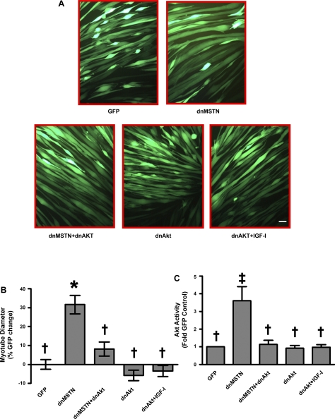
Inhibition of MSTN results in Akt-dependent myotube hypertrophy. A: representative photographs showing adenovirally infected, GFP-expressing myotubes. B: quantitation of mean myotube diameter ± SE expressed as percent GFP change (GFP, n = 104, dnMSTN, n = 129, dnMSTN+dnAkt n = 100, dnAkt, n = 42, dnAkt+IGF-I, n = 49). C: quantification of Akt kinase assay Western blots (data from n = 3 independent experiments for all except dnAkt, n = 4). *P < 0.001, ‡P < 0.01 vs. GFP, †P < 0.01 vs. dnMSTN; scale bar, 50 μm (magnification: 200).

