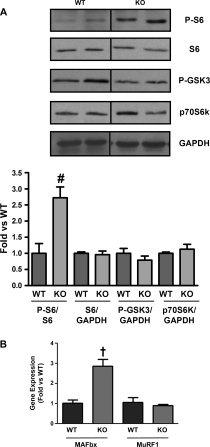
Fig. 6.
Increased S6 phosphorylation and atrogin-1/muscle atrophy F box (MAFbx) mRNA in vivo. A: representative Western blots from quadriceps of WT and KO mice was probed with anti-phospho-S6, S6, phospho-GSK3, p70S6K, and GAPDH antibodies. Bar graphs show quantification of blots normalized to S6 or GAPDH. Dividing lines on Western blot images depict where bands from the same blot have been juxtaposed. Quantitative real-time RT-PCR using RNA samples from WT and KO quadriceps shows an increase in MAFbx gene expression in KO skeletal muscle. Gene expression was normalized to GAPDH. (Western blots; n = 3WT:4KO). MURF-1, muscle RING-finger protein-1. Gene expression, n = 3WT:3KO. †P < 0.01 vs. WT, #P < 0.05 vs. WT.

