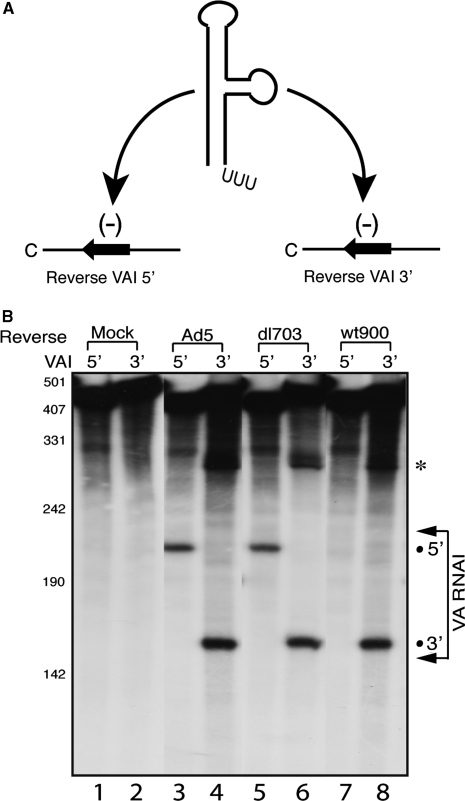
Figure 2.
The strand bias of mivaRNAI incorporating into RISC differs in wild-type Ad5, dl703 and wt900 infections. (A) The schematic diagram of RISC substrate RNAs. The VA RNAI gene was separated within the apical loop into two halves and cloned in anti sense orientation into a reporter mRNA. The 32P-labeled target RNAs containing the sequence complementary to the 5′ or 3′ halves of VA RNAI were generated by in vitro transcription. (B) S15 cytoplasmic extracts prepared from uninfected 293-Ago2 cells (Mock) or cells infected with wild-type Ad5, dl703 or wt900 were assayed for RISC activity against the reverse VAI 5′ or VAI 3′ target RNAs. Arrows indicate the span of the VA RNAI target regions in respective transcripts. The positions of cleavage products are indicated by dots. The bands labeled with ‘asterisks’ indicate the 3′-end cleavage products of the substrate RNAs, which is sometimes seen but usually degraded in S15 extracts.

